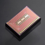
Tiny Injectable Sensor Could Provide Unobtrusive, Long-term Alcohol Monitoring
Engineers have developed a tiny, ultra-low power chip that could be injected just under the surface of the skin for continuous, long-term alcohol monitoring. The chip is powered wirelessly by a wearable device such as a smartwatch or patch. The goal of this work is to develop a convenient, routine monitoring device for patients in substance abuse treatment programs.


