Study Pinpoints Cellular Response to Pressure in Sea Star Embryos
Cells take up an unusual geometrical shape in response to pressure

Published Date
Story by:


Topics covered:
Share This:
Article Content
An international team of scientists has discovered a new cellular mechanism that explains how cells can adapt to pressure changes during tissue growth by packing themselves into a unique shape.
Researchers at UC San Diego’s Scripps Institution of Oceanography, Stanford University’s Hopkins Marine Station, and the Institute of Biomedicine in Seville (IBiS) in Spain led the research, which is novel for its use of sea star embryos as model organisms in this context. Their findings were published in the journal Development on May 7.
The lab work was conducted at Scripps Oceanography’s Center for Marine Biotechnology and Biomedicine (CMBB) in the Lyons Lab, which is focused on advancing the field of evolutionary developmental biology using marine invertebrates. The study is notable for its use of marine embryos — specifically the embryo of the sea star Patiria miniata — to understand how cells cope with changes in their physical environment.
“Our research shows that cells take up an unusual geometrical shape in response to pressure. It sheds light on how cells cope with changes in their physical environment, which happen dynamically in every tissue,” said lead author Vanessa Barone, who conducted the work while a postdoctoral researcher at Scripps Oceanography. “It is also a fascinating example of how studying a marine organism can lead to broadly relevant knowledge of fundamental cell biology.”
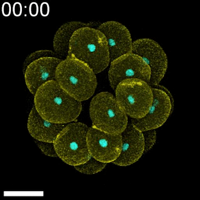
Timelapse of a developing sea star embryo, showing cell membranes in yellow and nuclei in cyan. Credit: Vanessa Barone
The authors said the results could have future implications for understanding how healthy cells could adapt to pressure exerted by tumor cells that grow uncontrollably.
While the cells’ unusual geometrical shape, a scutoid, had been described before, it was thought to occur mostly due to the shape of the tissue in which the cells are embedded. Scutoids have a prism-like shape, with six sides at the top and five sides at the bottom. Previous work has shown that when the tissue is curved in a certain way, like in tubes or egg-shaped forms, a proportion of the cells will become scutoids because that is the energetically favorable shape to have in that situation.
In the new study, the researchers used a combination of live imaging of sea star embryo development, detailed image analysis, and computational modeling to show that cells also become scutoids in other, much more common, circumstances.
They found that the cells became scutoids after cell divisions occurred in compact epithelial tissues. Cells are the building blocks of animals. During embryonic development, these cells rapidly divide, increasing in number. Epithelial cells distinguish themselves by their strong interconnections and ability to cover surfaces in the body. These cells form layers that create a protective barrier, separating external surfaces from internal cavities in adult animals. Additionally, epithelial tissue forms glands and is the predominant tissue in many organs, such as the liver or kidneys.
As the number of these cells increases, they often need to adjust to limited space, which leads to tissue compaction. Therefore, epithelial cells must organize themselves effectively, while withstanding the pressure from neighboring cells that are also proliferating. This study demonstrates that the epithelial cells were likely able to accommodate the newly formed cells by adopting a scutoid shape.
“By looking at the embryos of sea stars, we are uncovering important new information about cell biology, with potential connections to human health,” said Deirdre Lyons, a study co-author and marine biologist at Scripps Oceanography. “This is the first study to actually show the epithelial cell packing and cell division as the sea star embryo is developing, captured in live movies. Our findings have broad implications for understanding the cellular structure of these tissues.”
The sea star embryo is ideal for understanding how cells organize into an epithelial layer while they are proliferating. This is because sea star cells undergo several rounds of synchronous cell divisions that lead to the formation of an epithelial layer. Moreover, these embryos develop in seawater, they are fairly transparent and they are easy to image on a high-resolution microscope. These qualities enabled the scientists to follow each individual cell over time, while looking at the entire epithelial tissue as it forms.
"The proper coordination between cell growth and organization is a very complex process. By using the starfish embryo as a model, we have been able to dynamically study its early stages of development," said Luis María Escudero, a co-author of the study and researcher at IBiS.
The researchers at Scripps Oceanography captured live images in the lab showing these cell processes underway. The IBiS team then used CartoCell, a novel image analysis method recently published by Escudero's group, to further analyze the images. CartoCell is a deep-learning-based software tool that allows for quick and automatic processing of three-dimensional images, such as those from the sea star embryo timelapses.
“We observe that immediately after cell division, the probability of a cell adopting the scutoid shape increases significantly,” said Escudero. “Therefore, we conclude that the increase in cell density caused by proliferation is related to the change in shape. This change in shape probably occurs because cells better withstand compression when they are scutoids."
By demonstrating how cells organize within tissues in response to stress, this study could open the door to future applications related to cancer research.
“Our study could help in understanding the changes that occur in tissues that are compressed, whether because of normal processes or disease related situations,” said Barone, who is now an assistant professor at Stanford University.
In addition to Barone, Escudero, and Lyons, the research team included co-first author Antonio Tagua from IBiS, as well as study co-authors Jesus Á. Andrés-San Román and Juan Garrido-García from IBiS, and Amro Hamdoun from Scripps Oceanography.
The study was funded by the Human Frontier Science Program, the National Institutes of Health, and the Spanish Ministry of Science and Innovation.
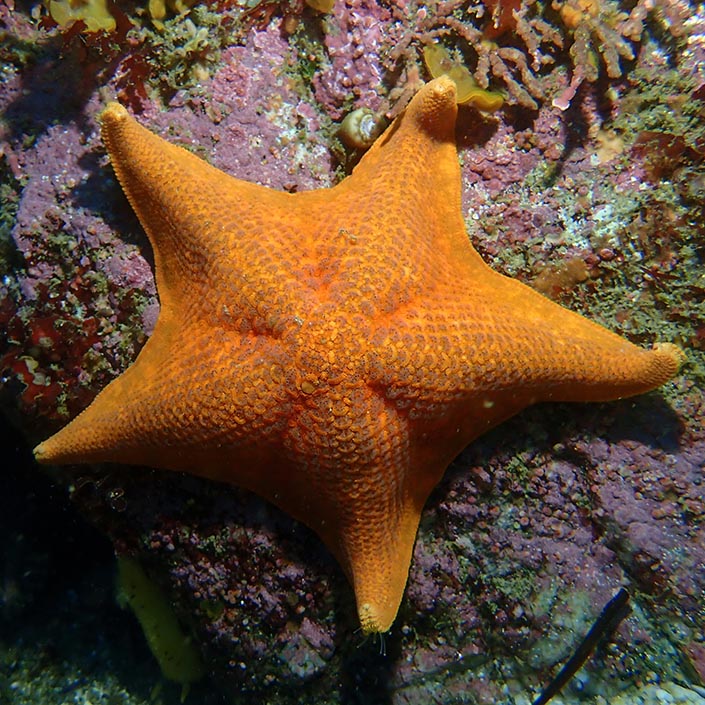
A bat star (Patiria miniata) observed in the wild off the Central Coast of California. Photo: Charlotte Seid
Topics covered:
Share This:
You May Also Like


Stay in the Know
Keep up with all the latest from UC San Diego. Subscribe to the newsletter today.