Following Cellular Lineage
Researchers present new findings on the development of the human forebrain
Story by:
Media contact:

Published Date
Story by:
Media contact:
Topics covered:
Share This:
Article Content
A group of researchers at University of California San Diego School of Medicine led an investigation that offers new insight into the development of the human forebrain.
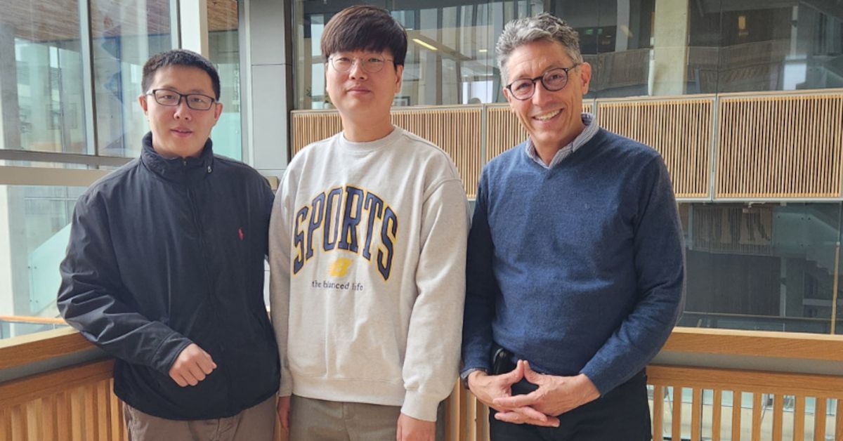
The study, led by Changuk Chung, Ph.D., and Xiaoxu Yang, Ph.D., both from the laboratory of Joseph G. Gleeson, M.D., Department of Neurosciences at the School of Medicine and the Rady Children’s Institute for Genomic Medicine, provide a greater understanding of how the human brain develops at the cellular level.
The study also presents evidence for the existence of the source of inhibitory neurons (dInNs) in the human brain that differ from origins in other species like mice, a common lab animal used in brain studies. The group outlined their findings in a paper recently published in the journal Nature.
The forebrain, or cerebral cortex, is the largest part of the brain, important for a wide range of function, ranging from cognitive thought, vision, attention and memory. Neurons are cells that serve as the individual circuits of the brain. Inhibitory neurons usually function as a kind of neural “off” switch, as opposed to the “on” switch of excitatory neurons.
“Humans have a very large and wrinkled cortex that likely supports higher cognitive functions compared with other species, such as rodents,” Gleeson explained.
He said that the inhibitory neurons in mice have an origin from deep within the developing brain. The current study puts that model to test by assessing cellular lineage. They found existence of dInNs, which are absent in mice. He said finding evidence for this specific type of neuron in humans opens the door to greater understanding how the human brain is special.
“We expect dInNs to support new, more accurate, models of human brains,” Gleeson said. “This updated brain model may help explain the origins of certain conditions like epilepsy, schizophrenia or autism.”
The group was especially interested in following the lineage trail of mosaic variants of brain cells. “If two cells share the same mother cell, we say they have the same lineage,” Chung said.
“If two individual cells have a same mosaic variant, they were born from a common mother cell that passed it to all of its daughters,” Yang explained. “So, mosaic variants in cells function like family names in people.”
The researchers directly accessed brains from two neurotypical donors who died from natural causes. They used mosaic variants to trace where these cells came from, to identify sister cells born in the same brain region, and to determine how far each “family name” spread across the brain.
They revealed that some inhibitory and excitatory neurons essentially have the same family name, which Chung said means the two types of neurons share lineage. The two types likely branched in a late moment of embryonic cerebral development, he added, noting that such a cellular relationship is not present in other species.
Presentations by the UC San Diego forebrain researchers featured this AI image of the brain, generated by LLM through ChatGTP4.
“We hope our paper helps other researchers generate better models of neurological disease, and which types of brain diseases can result from impaired development,” Gleeson concluded.
In addition to Changuk Chung, Xiaoxu Yang and Joseph G. Gleeson, co-authors at University of California San Diego are: Robert F. Hevner, Sanford Consortium for Regenerative Medicine, School of Medicine Department of Pathology; Keng Ioi Vong, School of Medicine Department of Neurosciences, and Rady Children’s Institute for Genomic Medicine; Yang Liu, School of Medicine Department of Neurosciences and Rady Children’s Institute for Genomic Medicine; Arzoo Patel, School of Medicine Department of Neurosciences, and Rady Children’s Institute for Genomic Medicine; Rahul Nedunuri, School of Medicine Department of Neurosciences, and Rady Children’s Institute for Genomic Medicine; Scott T. Barton, School of Medicine Division of Medical Education; Geoffroy Noel, School of Medicine Division of Anatomy; Chelsea Barrows, School of Medicine Department of Neurosciences, and Rady Children’s Institute for Genomic Medicine; Valentina Stanley, School of Medicine Department of Neurosciences, and Rady Children’s Institute for Genomic Medicine; Swapnil Mittal, School of Medicine Department of Neurosciences, and Rady Children’s Institute for Genomic Medicine; Johannes C.M. Schlachetzki, School of Medicine Department of Neurosciences, and Department of Cellular and Molecular Medicine.
Other co-authors are: Stephen F. Kingsmore, Rady Children’s Institute for Genomic Medicine; Katie Kennedy, BioSkryb Genomics Inc.; Martin W. Breuss, Department of Pediatrics, Section of Clinical Genetics and Metabolism, University of Colorado Aurora.
This work was supported by National Institute of Mental Health (NIMH) grants U01MH108898, R01MH124890 and R21MH134401; a Larry L. Hillblom Foundation Grant; a Eunice Kennedy Shriver National Institute of Child Health and Human Development (NICHD) grant K99HD111686; a 2021 NARSAD Young Investigator Grant from the Brain & Behavior Research Foundation; and the Rady Children’s Institute for Genomic Medicine.
Competing interests: All authors declare no competing interests.
Read more news about: School of Medicine, Health Sciences
“This updated brain model may help explain the origins of certain conditions like epilepsy, schizophrenia or autism.”


Stay in the Know
Keep up with all the latest from UC San Diego. Subscribe to the newsletter today.