Research Alerts
Understanding the Mechanisms of Embryonic Cell Behavior


Published Date
Article Content
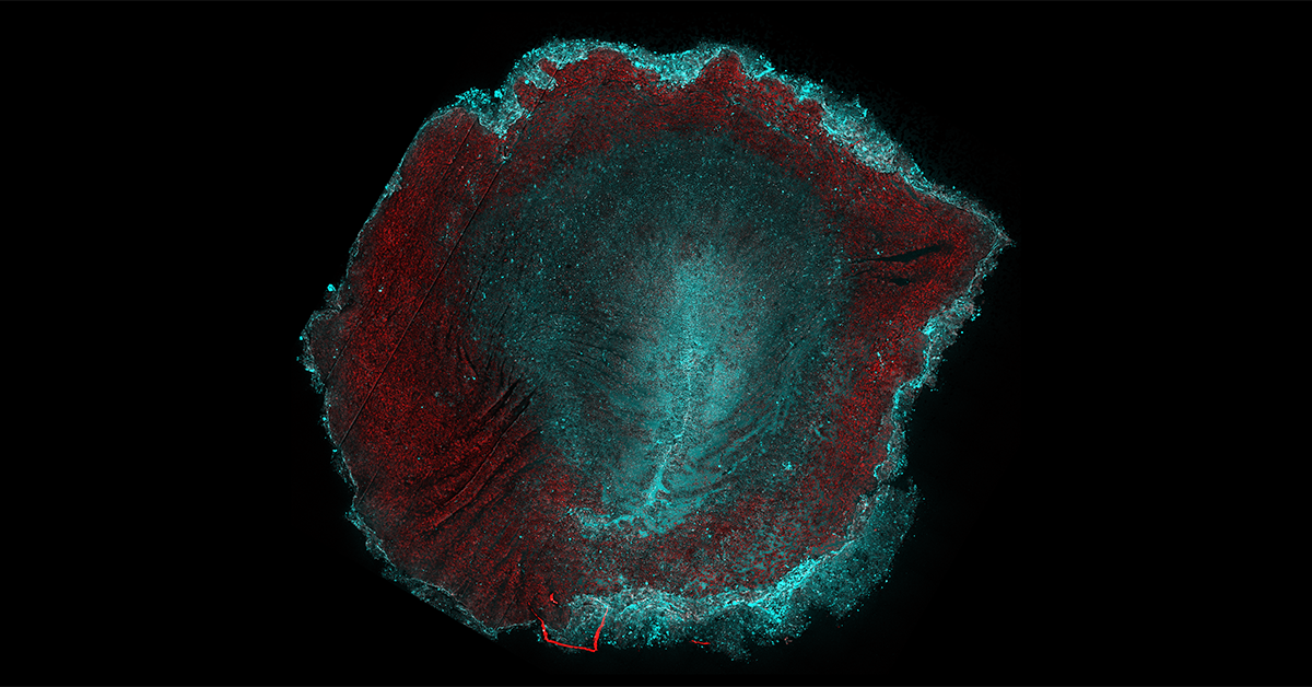
During embryonic development, thousands of cells divide and move collectively, sculpting the main body axes from an initially symmetric ensemble of cells. Understanding the mechanisms that coordinate this collective behavior remains a significant challenge in biology and the physics of living systems, but a better understanding could have implications in health and science, from medicine to biomaterials.
Now researchers have discovered that avian embryos — established models for studying human development — control their size and shape using modular, independent physical mechanisms.
Clarifying the modular mechanisms that regulate the emergent embryo geometry (size and shape) helps further our understanding of the evolutionary plasticity of natural embryos and suggests strategies for engineering synthetic ones.
The study, published June 4, 2025 in Nature Communications, was led by UC San Diego researchers Alex Plum and Mattia Serra, along with Guillermo Serrano Najera and Ben Steventon (both University of Cambridge) and Kees Weijer (University of Dundee). Their research was funded by the National Sciences Foundation (PHY-2413073 and CAREER PHY-2443851).
Read the study in Nature Communications: Control of Tissue Flows and Embryo Geometry in Avian Gastrulation.
Read more news about: Physical Sciences
“Physical principles offer remarkable insight into how similar cells self-organize into complex, functional forms.”
Share This:


Stay in the Know
Keep up with all the latest from UC San Diego. Subscribe to the newsletter today.