UC San Diego Researchers Awarded $2 Million from NIH to Visualize Human Brain’s Magnetic Activity
New magnetoencephalography center opens at the UC San Diego Qualcomm Institute this fall
Story by:
Published Date
Article Content
The National Institutes of Health (NIH) has awarded $2 million to a UC San Diego research center at the Qualcomm Institute (QI) for the purchase of a new, non-invasive magnetoencephalography (MEG) system to study the magnetic fields of the human brain. The research has applications for disorders such as epilepsy, brain tumors, concussions, schizophrenia, post-traumatic stress disorder, autism spectrum disorder and Alzheimer’s disease.
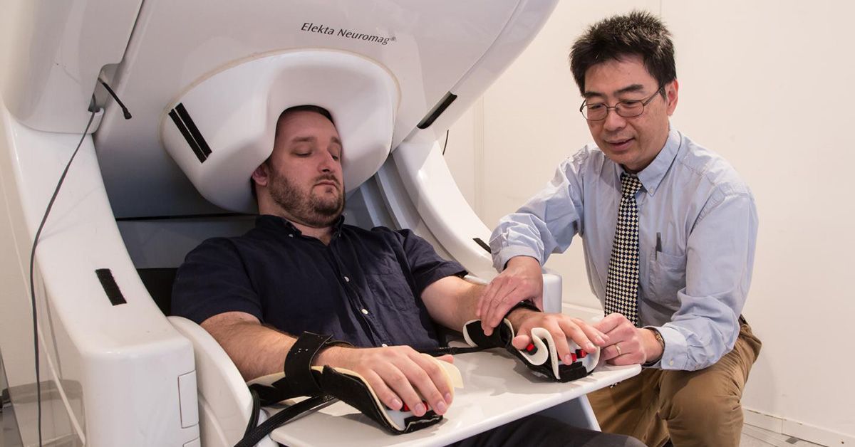
Roland Lee, M.D., director of the MEG Center at QI, and Mingxiong Huang, Ph.D., center co-director, will use the funds to secure a state-of-the-art MEG machine that can help researchers visualize the brain’s activity by measuring the magnetic fields generated by electrical currents from brain cells (neurons) as they fire in real-time. The MEG can aid scientists and physicians in pinpointing seizure sites, mapping brain function ahead of surgery and developing imaging markers for aiding in the diagnosis of brain disorders.
The MEG will sit on the first floor of QI’s Atkinson Hall on the UC San Diego campus, ensconced within a room-sized insulated chamber shielded from outside electromagnetic interference by six layers of metal. The only one of its kind in Southern California, the machine will support the lab’s research as well as be widely available as a shared service to other scientists and to medical patients.
“This new state-of-art MEG instrument replaces our old MEG unit installed in 2005, and will have improved signal and faster electronics,” said Lee, who is also emeritus professor of radiology at UC San Diego School of Medicine and a physician with the Veterans Affairs (VA) San Diego Healthcare System. “We will continue to serve as the UC San Diego core MEG facility for research scientists, and physicians and their patients throughout southern California and the Southwestern U.S.”
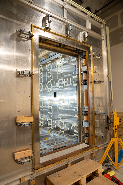
The insulated chamber that will house the new MEG machine in QI’s Atkinson Hall. Photo by Alex Matthews/QI.
A Better Way to “See” the Brain Work
When a person thinks, acts or experiences an emotion, their neurons fire up and create an electrical current. In turn, this current gives rise to electric and weak magnetic fields that can be detected using modern devices, including MEG.
While MRIs, electroencephalograms (EEGs) and other technologies can also provide a look inside the brain, the MEG allows researchers and clinicians to detect brain activity in real-time and in much finer detail than an EEG. Users can pinpoint the site of neuronal activity within three millimeters and in mere milliseconds, whereas functional MRI, which measures small amounts of blood flow demanded by active brain regions but does not measure brain cell activity, though spatially accurate, lags seconds behind.
Unlike an EEG, a MEG has an advantage in that it measures magnetic fields instead of electrical fields. As they pass through the human skull and tissues, electrical fields can easily become muffled or distorted, a fact that makes it difficult for researchers to calculate where in the brain they originate. Because the only thing that stops magnetic fields is metal, MEG can provide a clear, uninterrupted signal using a helmet equipped with sensors—rather than surgically implanted electrodes on the brain’s surface.
“The noninvasive component to MEG is really important,” says Huang, who is also affiliated with the Department of Electrical and Computer Engineering at the UC San Diego Jacobs School of Engineering and the Department of Radiology at UC San Diego School of Medicine, as well as the VA San Diego Healthcare System.
When Patients Need Help
When might a researcher or clinician need this level of information about brain function? Take the example of the nearly 30 to 40% cases of epilepsy that do not respond to medication. In these more severe cases, Lee says, clinicians need to be able to clearly identify the part of the brain causing a misfire to determine whether surgery via laser or scalpel is a viable treatment option. MEG is ideally suited to provide this information.
Tumors can also distort the surface and appearance of the brain to such a degree that important regions, like those used in movement and language processing, become difficult to locate. MEG data can help surgeons plan their operations with more information regarding locations of this important functioning brain tissue before performing surgery.
Concussions are another application. Unlike the most catastrophic cases of brain trauma, which show up on MRI and CT scans, concussions have been elusive to diagnose. MEG provides a new tool on that front, by showing injured brain regions which demonstrate abnormally slow brain magnetic rhythms, even though these regions appear normal on CT and MRI. “We can also use the MEG to follow patient recovery, including in cases with repeated concussions,” notes Lee.
For more information on using the equipment at the MEG Center, see https://qi.ucsd.edu/services/high-tech-research/meg-center/. The MEG will open to clinicians and scientists regardless of affiliation with the university this fall.
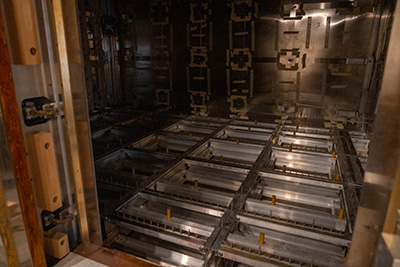
A view inside the MEG’s new insulated chamber, before the final installation of its floors and walls. Photo by Alex Matthews/QI.
Lee and Huang commented: “We are grateful to QI Director Ramesh Rao and his team for their strong support of the MEG Center. We are also grateful for the contributions of colleagues from more than 20 UC San Diego research groups with projects funded by the NIH, the U.S. Departments of Veterans Affairs and Defense, and other funding agencies. All of these groups contributed to the success of this award.”
Share This:
You May Also Like


Stay in the Know
Keep up with all the latest from UC San Diego. Subscribe to the newsletter today.