Roger Tsien Inducted Into National Inventors Hall of Fame
With two others, the late professor of chemistry won the Nobel Prize in 2008 for creating glowing proteins that can illuminate the internal machinery and processes of life
Story by:
Published Date
Article Content
In life, Roger Tsien quite literally helped illuminate it, creating a palette of glowing proteins that could be used to peer within living cells and observe functions never seen before. The achievement, with Osamu Shimomura and Martin Chalfie, earned the trio the 2008 Nobel Prize in chemistry.
Tsien, who was a professor in the Department of Pharmacology, Chemistry and Biochemistry at UC San Diego School of Medicine, died in 2016 at the age of 64, but celebrations of his enduring contributions to science continue.
On Friday, January 6, the National Inventors Hall of Fame (NIHF) announced Tsien would be inducted as a member of the 2023 class, along with inventors of CRISPR-Cas9 gene editing, the mRNA technology underpinning COVID-19 vaccines, a type of stainless steel, new wheelchair technologies and reCAPTCHA, the ubiquitous website security system deployed to distinguish between human and automated users.
The 16 inductees are part of the NIHF’s 50th anniversary, which was founded in 1973 when Thomas Edison was the sole inductee.
Tsien, Shimomura, now an emeritus professor at the Marine Biological Laboratory in Woods Hole, Mass. and Martin Chalfie, PhD, a professor of biological sciences at Columbia University, collaborated to discover and develop green fluorescent protein (GFP), derived from the jellyfish Aequorea victoria, as a new and soon-indispensable research tool.
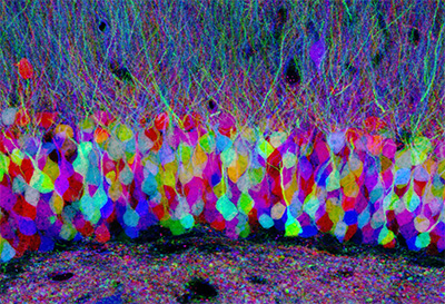
“Brainbow” mice are engineered with a gene that includes three different fluorescent proteins, though only one color is actually expressed from each of DNA construct. In this micrograph of a mouse hippocampus, neurons randomly choose combinations of red, yellow and cyan fluorescent proteins. The colors provide a way to distinguish neighboring neurons and visualize brain circuits. Cell Image Library
Shimomura identified the crucial jellyfish protein and revealed that it glowed bright green under ultraviolet light. Chalfie showed how it could be used as a biological marker. Combining his deep skills in chemistry and biology, Tsien found ways to make GFP glow more brightly and consistently; then he created a full palette of fluorescent proteins that scientists could use to track different cellular processes at the same time.
“I’ve always been attracted to colors,” Tsien told The San Diego Union-Tribune in 2008. “Color helps make the work more interesting and endurable. It helps when things aren’t going well. If I had been born color-blind, I probably never would have gone into this.”
The utility of GFP proteins was immediately and broadly recognized, making it possible for biologists to mark and track molecules and functions within living cells in real-time. It became possible, for example, to monitor DNA and which proteins were used to perform different duties; audit gene expression in different kinds of cells and tag molecules as they move in and out of cells.
Emerging GFP tools did not stay locked within laboratories. In 2010, Tsien, Quyen T. Nguyen, MD, PhD, professor of Surgery at UC San Diego School of Medicine and a head-and-neck surgeon at UC San Diego Health and others described using fluorescent probes to light up cancerous tumors. A year later, they published findings about using GFP in surgery, injecting fluorescent peptides into mouse models that caused hard-to-see peripheral nerves to glow, alerting surgeons to their location even before the nerves are encountered.
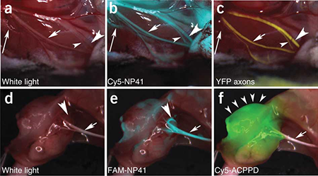
Quyen Nguyen, Tsien and others showed in 2011 how fluorescent proteins injected into mouse tissues could be used to light up critical structures, such as nerves, that would be invisible under normal light. Photo by UC San Diego Health
In 2019, UC San Diego Health opened the Center for Fluorescence-Guided Surgery to leverage the rapidly evolving imaging tool across a spectrum of procedures, from common gallbladder removal and weight loss surgery to complex neurosurgical operations.
Perhaps the greatest potential lies in the drugs that Tsien co-invented using fluorescence to help surgeons ensure safe and complete removal of malignant tissues while preserving critical nerves during surgery.
For example, pegloprastide (licensed from UC San Diego by Avelas Biosciences) is a fluorescent imaging agent currently in clinical trials for patients undergoing breast cancer detection. It received “breakthrough therapy” designation from the U.S. Food and Drug Administration (FDA) in 2020. Bevonescein (licensed from UC San Diego by Alume Biosciences) is another fluorescent marker in clinical trials designed to locate nerves during head and neck surgeries. It recently received FDA fast track designation intended to speed development.
“These technologies are legacies of Roger's desire to use fluorescence to make a difference in patient outcomes. Both are in the late stages of development and have received significant FDA recognition for their high clinical impact potential,” said Nguyen. “Roger was a visionary who saw the power and possibilities of fluorescence. His work continues to light the way.”
The smaller picture
Tsien did not rest on his Nobel laurels.
In 2011, he and colleagues created a new type of genetic tag visible under an electron microscope (EM), providing even greater cellular detail.
“The big advantage of EM is that it has always had much higher spatial resolution than light microscopy. You can get up to a hundred-fold higher useful magnification from EM than from light microscopy,” said Tsien at the time.
“Existing EM images of cell structures are analogous to maps or aerial photographs showing major landmarks and populations centers. It was very difficult to see where individual protein types were located, just as most geographical maps do not show the location of individual classes of people, such as everyone with the surname Smith, or all of the orthodontists in an area. Our new technique enables us to put beacons on just about any protein and get a snapshot of its location at the much higher resolution of EM.”
Tsien studiously avoided the spotlight, but scores of awards and accolades found him.
“Every honor was justly deserved, and always received with humility,” said Pradeep Khosla, chancellor of UC San Diego when Tsien died. “Roger was an extraordinary man: kind, generous, gracious, and always the consummate scientist pushing the limits of his work to expand the possibilities of science. He was a rare talent we cannot replace.”
His achievements, though, shine on.
You May Also Like


Engineers Take a Closer Look at How a Plant Virus Primes the Immune System to Fight Cancer
Technology & EngineeringStay in the Know
Keep up with all the latest from UC San Diego. Subscribe to the newsletter today.