Appendiceal Cancer Gets Its Own Preclinical Model
UC San Diego researchers develop a 3-D culture for a rare disease that has long defied effective study and development of therapies
Story by:
Published Date
Article Content
Appendiceal cancer (malignancies of the appendix, a small tissue pouch that is part of the gastrointestinal tract) is very rare, occurring in perhaps one or two people per 1 million per year. Prognoses is mixed, with a 5-year survival rate of 67 to 97 percent for low-grade tumors detected early, but much lower for advanced cases that may have spread to other parts of the body.
For cancer researchers attempting to study appendiceal cancer and find remedies, a primary challenge has been the lack of an effective preclinical model to probe its pathology and test new drugs or therapeutic approaches.

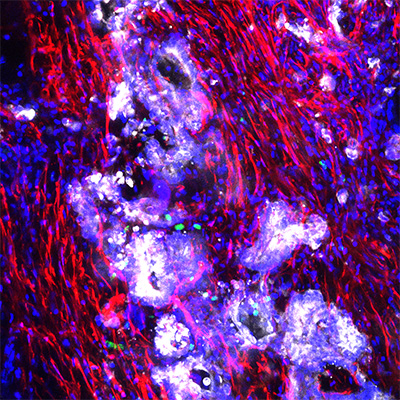
In a paper published in the November 1, 2022 print issue of Clinical Cancer Research, researchers at University of California San Diego School of Medicine and Moores Cancer Center at UC San Diego Health describe the first preclinical model of appendiceal cancer that contains all elements of the tumor, allowing previously stymied investigations to proceed.
“We’ve learned that appendiceal cancer has a distinctive genomic landscape and is surprisingly full of immune cells,” said senior author Andrew Lowy, MD, chief of the Division of Surgical Oncology at Moores Cancer Center at UC San Diego Health and a professor of surgery at UC San Diego School of Medicine.
”Relying on other models, such as colorectal, don’t apply, which makes this an unmet need. Epithelial neoplasms (new, abnormal tissue growth) of the appendix are rare, but without an effective way to study them, the opportunities to develop new treatments have also been rare.”
The obstacles to creating a preclinical model of appendiceal cancer are numerous:
- Access to clinical tissues is rare because the disease is rare.
- The majority of neoplasms have mucinous histology, a characteristic that makes them difficult to assess under a microscope and to culture.
- Mice do not have the equivalent of a human appendix, rendering them unsuitable as a genetically modified model.
Lowy and colleagues developed an organotypic slice culture of living appendiceal cancer cells. Organotypic slices are three-dimensional cultures of an organ. In this case, the slices are made from appendiceal cancer tissue removed from patients at surgery, then cultured ex vivo, or outside, of the patient. Organotypic slices have been created to model other cancers, such as pancreatic, lung and colon, but not appendiceal, until now.
“We’ve learned that appendiceal cancer has a distinctive genomic landscape and is surprisingly full of immune cells.”

The authors said the organotypic model accurately recapitulates appendiceal tumors in terms of cell mix and behavior, which makes it a promising new research tool in addition to other approaches, such as organoids and patient-derived xenografts (transplanted tissue grown in another species, such as a mouse or rat).
“We’ve learned that utilizing tissue slices made from patient tumor resections is great way to study the pathobiology of this disease, and we are hopeful they will help predict therapy responses in patients,” said Lowy. “Using this new model, we can now test new treatments that might lead to better outcomes for patients with advanced disease.”
Co-authors include: Jonathan Weitz, Tatiana Hurtado de Mendoza, Herve Tiriac, James Lee, Siming Sun, Bharti Garg, Jay Patel, Kevin Li, Joel Baumgartner, Kaitlin J. Kelly, Jula Veerapong, Morgan Hossein and Yuan Chen, all at UC San Diego.
Funding for this research came, in part, from the National Organization for Rare Disorders (grants R21CA273974, 1F32CA265052-01) and gifts from the estate of Elisabeth and Ad Creemers, the Euske Family Foundation, the Gastrointestinal Cancer Research Fund and the Peritoneal Metastasis Research Fund.
Stay in the Know
Keep up with all the latest from UC San Diego. Subscribe to the newsletter today.


