Evolution of Kangaroo-Like Jerboas Sheds Light on Limb Development
Published Date
By:
- Kim McDonald
Share This:
Article Content

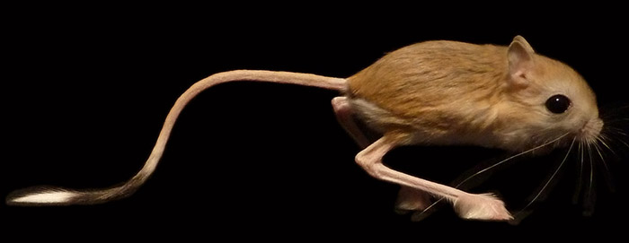
Lesser Egyptian Jerboa. Photo by Cooper Lab, UC San Diego
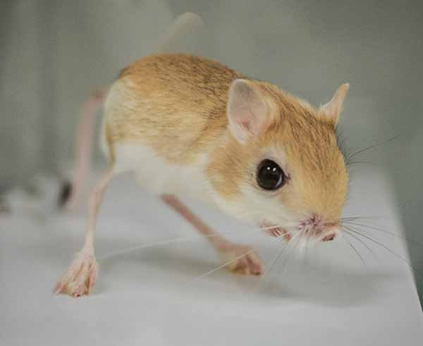
With their tiny forelimbs and long hindlimbs and feet, jerboas are oddly proportioned creatures that look something like a pint-size cross between a kangaroo and the common mouse.
How these 33 species of desert-dwelling rodents from Northern Africa and Asia evolved their remarkable limbs over the past 50 million years from a five-toed, quadrupedal ancestor shared with the modern mouse to the three-toed bipedal jerboa is detailed in a paper published in this week’s issue of the journal Current Biology.
The study—which examines how natural selection altered limb structure and biomechanics in the jerboa—is more than a detailed picture of the evolution of limb and digit development in this curious-looking rodent. It also sets the stage to understand processes that control the development of limbs and the remodeling of bones for medical scientists looking for new ways to treat osteoporosis and abnormalities in human limb development.
“When the human embryo starts to form the humerus, radius, ulna and the digits of the hand, they initially form at the same size, but the rates of growth of each of those bones change during development,” explained Kimberly Cooper, an assistant professor of biology at UC San Diego who headed the study. “How that happens, how we get a difference in growth so that the smaller bones grow at a slower rate for a shorter period of time than the long bones is something we don’t understand. To answer these questions, we have been studying the jerboa, which has evolved dramatically different skeletal proportions from the mouse, such as elongated hind legs, particularly the feet. This disproportionality has given us a window into understanding the mechanisms of how proportions are established in humans and other vertebrates.”
Scientists believe jerboas evolved when the Tibetan plateau began changing 50 million years ago to a more arid climate, reducing vegetation—the natural hiding places for small rodents—and requiring them to travel long distances to search for seeds and other plant food.
“One advantage that jerboas have is that they can travel long distances and avoid predators really easily because they are bipedal,” said Cooper. “Jerboas can change direction rapidly and are capable of jumping as far as six feet and three feet straight in the air.”
By lengthening the hindlimb and reducing the muscle mass and number of toes in their feet, as well as by fusing the long bones of their hind paws, jerboas evolved to become more mechanically efficient as runners. With less mass to swing at the end of their legs, they could travel for longer distances across the desert in search of food or places to hide.
“The evolution of horses is a great comparison,” explained Cooper. “In the horse, digits were lost, foot muscles were reduced and limbs were elongated. All of those things increased the mechanical advantage in locomotion. When you lengthen the foot, you can have a longer stride. Digit and muscle loss makes it easier mechanically to move. That’s what happened in both the jerboas and horses.”
To answer the question of how evolution and developmental biology worked together to increase hindlimb proportions, decrease the digits and fuse the foot bones, a team of scientists from Harvard University and Montana State University worked together with Cooper over the past seven years to detail the skeletal anatomy of the 33 species of jerboas from museums around the world and compare them to closely related species of quadrupedal mice, such as the woodland jumping mouse and the birch mouse.

“There is enormous diversity in the proportions of limbs between the species,” said Cooper. “One goal was to determine the genetic complexity of the mechanisms that established the proportions of the forelimb versus the hindlimbs, as well as the mechanisms that established proportions within a limb.”
The scientists found that many of those processes worked independently. Because there are species of jerboas that have longer hindlimbs that don’t necessarily have longer feet, for instance, the process of producing a longer hindlimb does not always result in a longer foot.
“We found that evolution controls individual bone size,” said Cooper. “That’s huge, because if you think about the genes that control the growth of all of the different bones in our body, that suggests they have to be regulated in very complex ways.”
Finding those genetic regulatory factors, which Cooper hopes to pursue in a future research effort, could help medical researchers develop ways to control growth asymmetries in human limb development. The current method of correcting bone asymmetries is a process called distraction osteogenesis, in which a bone is broken in two and separated to induce it to lay down new bone and lengthen. But that involves major and costly surgery.
“If we could identify the local factors in growth and deliver it with a pump locally to the growth plate, we could have a more precise and less invasive way of correcting limb proportion in juveniles,” said Cooper.
Cooper and her colleagues also plan to study in more detail the cellular processes by which parallel bones fuse in the jerboa foot. Two cell types work together to make that happen: osteoclasts, which chew up bone, and osteoblasts, which lay down new bone.
“This process occurs normally in all of us, and osteoporosis is when you have an imbalance—you have less osteoblast activity and more osteoclast activity, so you chew up bone, but you don’t remake it at the same rate,” Cooper explained. “We tend to think of these two cell types working together, but in the juvenile jerboa they are geographically in different locations, because you’ve got bone being laid down in one place and bone being chewed up in another place.”
How these two cell types communicate with one another to regulate this process in two different locations should provide some insights useful to the treatment of osteoporosis.
“We want to understand how you get an uncoupling of the activities of these two cell types so they can each do their job, but in two different locations within the skeletal element,” she said. “If we can understand more about the communication of these two cell types, we may be able to have a major impact on osteoporosis.”
The other co-authors of the paper were Talia Moore, Scott Edwards, Andrew Biewener, Farish Jenkins and Clifford Tabin of Harvard University and Chris Organ of Montana State University.
Share This:
You May Also Like
Stay in the Know
Keep up with all the latest from UC San Diego. Subscribe to the newsletter today.


