Wearable Ultrasound Patch Enables Continuous, Non-Invasive Monitoring of Cerebral Blood Flow
Story by:
Published Date
Story by:
Topics covered:
Share This:
Article Content
Engineers at the University of California San Diego have developed a wearable ultrasound patch that can offer continuous, non-invasive monitoring of blood flow in the brain. The soft and stretchy patch can be comfortably worn on the temple to provide three-dimensional data on cerebral blood flow—a first in wearable technology.
A team of researchers led by Sheng Xu, a professor in the Aiiso Yufeng Li Family Department of Chemical and Nano Engineering at the UC San Diego Jacobs School of Engineering, published their new technology on May 22 in Nature.
The wearable ultrasound patch marks a significant leap from the current clinical standard, called transcranial Doppler ultrasound. This method requires a trained technician to hold an ultrasound probe against a patient’s head. The process has its downsides, however. It is operator-dependent, so the accuracy of the measurement can vary based on the operator’s skill. It is also impractical for long-term use.
Xu’s team developed a device that overcomes these hurdles. Their wearable ultrasound patch offers a hands-free, consistent and comfortable solution that can be worn continuously during a patient’s hospital stay.
“The continuous monitoring capability of the patch addresses a critical gap in current clinical practices,” said study co-first author Sai Zhou, a materials science and engineering Ph.D. candidate in Xu’s lab. “Typically, cerebral blood flow is monitored at specific times each day, and those measurements do not necessarily reflect what may happen during the rest of the day. There can be undetected fluctuations between measurements. If a patient is about to experience an onset of stroke in the middle of the night, this device could offer information that is crucial for timely intervention.”
Patients who are undergoing and recovering from brain surgery can also benefit from this technology, noted Geonho Park, another co-first author of this study who is a chemical and nano engineering Ph.D. student in Xu’s lab.
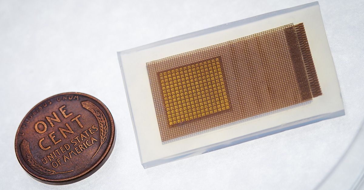
The patch, roughly the size of a postage stamp, is constructed from a silicone elastomer embedded with several layers of stretchy electronics. One layer consists of an array of small piezoelectric transducers, which produce ultrasound waves when electrically stimulated and receive ultrasound waves reflected from the brain. Another key component is a copper mesh layer—made of spring-shaped wires—that enhances signal quality by minimizing interference from the wearer’s body and environment. The rest of the layers consist of stretchable electrodes.
During use, the patch is connected through cables to a power source and computer. To achieve 3D monitoring, the researchers integrated ultrafast ultrasound imaging into the system. Unlike standard ultrasound, which captures about 30 images per second, ultrafast imaging captures thousands of images per second. This high frame rate is necessary for collecting robust data from the piezoelectric transducers in the patch, which would otherwise suffer from low signal intensity due to the strong reflection of the skull.
The data are then post-processed using custom algorithms to reconstruct 3D information such as the size, angle and position of the brain’s major arteries.
“The cerebral vasculature is a complex structure with multiple branching vessels. You need a device capable of capturing this three-dimensional information to get the whole picture and obtain more accurate measurements,” said Xinyi Yang, another co-first author of this study and materials science and engineering Ph.D. student in Xu’s lab.
In this study, the patch was tested on 36 healthy volunteers for its ability to measure blood flow velocities—peak systolic, mean flow and end diastolic velocities—in the brain’s major arteries. Participants engaged in activities affecting blood flow, such as hand-gripping, breath-holding and reading. The patch’s measurements closely matched those obtained with a conventional ultrasound probe.
Next, the researchers plan to collaborate with clinicians at UC San Diego School of Medicine to test the patch on patients with neurological conditions that impact cerebral blood flow. Xu has co-founded a startup company called Softsonics to commercialize this technology.
Paper: “Transcranial volumetric imaging using a conformal ultrasound patch.”
This work was supported by the National Institutes of Health (1R21EB025521-01, 1R21EB027303-01A1, 3R21EB027303-02S1, 1R01EB033464-01, 1R01HL171652-01).

Sai Zhou, co-first author of the study and UC San Diego materials science and engineering Ph.D. candidate.


Stay in the Know
Keep up with all the latest from UC San Diego. Subscribe to the newsletter today.