Butterfly Wings Inspire New Imaging Technique for Cancer Diagnosis
Using the nanostructures found on butterfly wings, scientists have developed a simple and inexpensive way to analyze cancerous tissues.
Story by:
Published Date
Story by:
Topics covered:
Share This:
Article Content
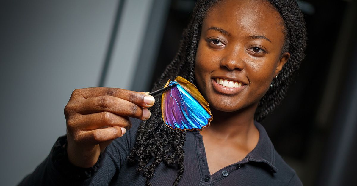
Researchers at the University of California San Diego have found an unusual ally in the quest to make cancer diagnosis faster, more accurate and more accessible worldwide: the Morpho butterfly. Known for its shimmering blue wings, the Morpho butterfly owes its brilliance not to pigments but to microscopic structures that manipulate light. Now, researchers are harnessing those same structures to gain detailed insights into the fibrous makeup of cancer biopsy samples—without the need for chemical staining or expensive imaging equipment.
The findings are detailed in a paper published in Advanced Materials.
Fibrosis, the accumulation of fibrous tissue, is a key feature of many diseases, including neurodegenerative disorders, heart disease and cancer. In oncology, evaluating the extent of fibrosis in a biopsy sample can help determine whether a patient’s cancer is in an early or advanced stage.
“The big challenge, however, is that it is extremely difficult to distinguish between these stages using current clinical methods,” said study senior author Lisa Poulikakos, a professor in the Department of Mechanical and Aerospace Engineering at the UC San Diego Jacobs School of Engineering. These methods rely on staining tissues to highlight key structures in the tumor biopsy, but the results can be subjective—one pathologist might interpret a sample differently from another. And while there are more advanced imaging techniques that can provide richer detail, they require expensive, specialized equipment that many clinics simply don’t have.
That’s where the Morpho butterfly comes in. Poulikakos and her team discovered that by placing a biopsy sample on top of a Morpho butterfly wing and viewing it under a standard microscope, they can assess whether a tumor’s structure indicates early- or late-stage cancer—without the need for stains or costly imaging machines.
“We can apply this technique using standard optical microscopes that clinics already have,” said Poulikakos. “And it’s more objective and quantitative than what is currently available.”
The idea for this method came from Paula Kirya, a mechanical engineering graduate student at UC San Diego and the study’s first author. Kirya had previously studied the Morpho butterfly’s wings and their optical properties while an undergraduate student researcher at Pasadena City College. When she transferred to UC San Diego and joined Poulikakos’ lab—where researchers build synthetic nanostructures to image biological tissues—she recognized an opportunity.
“I had been imaging butterfly wings, studying how they react to different environments,” she said. “And when I saw what the lab was doing, I thought, ‘The Morpho naturally has this property—why not use it?’”
The researchers found that the wing’s micro- and nanostructures respond strongly to polarized light—a kind of light that propagates in a specific direction. Collagen fibers—which are a key structural component of fibrotic tissue—also interact with polarized light, but their signals are weak. By placing a biopsy sample above a piece of a Morpho butterfly wing, the researchers amplified these signals, making it easier to analyze the density and arrangement of collagen fibers.
The resulting signals can then be translated into a measure of just how dense and organized the collagen fibers are in the biopsy sample. To do this, the researchers developed a mathematical model based on Jones calculus, a method for analyzing polarized light. The model correlates light intensity with the density and organization of collagen fibers, providing a quantifiable metric to assess fibrosis within the tissue.

Lisa Poulikakos
Using this approach, the researchers analyzed both collagen-dense and collagen-sparse human breast cancer biopsy samples provided by study collaborators and co-authors Jing Yang, a professor in the Departments of Pharmacology and Pediatrics at UC San Diego School of Medicine and co-leader of the Cancer Biology and Signaling Program at Moores Cancer Center, and Aida Mestre-Farrera, a postdoctoral scientist in Yang’s group. Their results were comparable to conventional staining methods and an advanced, high-cost imaging method.
“Essentially, we’re trying to expand on these procedures with a stain-free alternative that requires nothing more than a standard optical microscope and a piece of a Morpho wing,” said Kirya. “In many parts of the world, early cancer screening is a challenge because of resource limitations. If we can provide a simpler and more accessible tool, we can help more patients get diagnosed before their cancers reach aggressive stages.”
While the current study focused on breast cancer, the researchers believe their technique could be applied to a wide range of fibrotic diseases.
“We’re excited to leverage this technique for all kinds of tissue diagnostics,” said Poulikakos. “It was really surprising to see how well nature had already designed a solution via the Morpho butterfly wing and its natural micro- and nanostructures. Our work shows that nature has given us something that can help us image diseased tissues without the need for expensive fabrication facilities.”
This work was supported by the National Cancer Institute (R01CA174869, RO1CA262794, R01CA268179 and R01CA236386), a Beckman Young Investigator award by the Arnold and Mabel Beckman Foundation (Project Number: 30155266), an Optica Amplify Scholarship, an Optica Women’s Scholarship, a Selected Professions Fellowship from the American Association of University Women, the National Science Foundation Graduate Research Fellowship Program (DGE-2038238), a Krueger v. Wyeth research award, and a TRDRP Postdoctoral Award (T32FT4922).

Jing Yang (left) and Aida Mestre-Farrera (right)


Stay in the Know
Keep up with all the latest from UC San Diego. Subscribe to the newsletter today.