

Bioengineers Awarded $14M from NIH to Build Digital Maps of Brain, Other Organs at Single-Cell Level

Published Date
By:
- Liezel Labios
Share This:
Article Content
Kun Zhang, professor of bioengineering at the University of California San Diego Jacobs School of Engineering, has received $14 million in grants from the National Institutes of Health to build 3D, digital single-cell maps of the human brain and organs in the respiratory and urinary systems.
The work aims to provide a deeper understanding of the functions and malfunctions of organs in the human body at the level of individual cells.
“We’re building so-called ‘reference maps’ for these organs. For example, if a patient has Alzheimer’s or chronic kidney disease, we can zoom in and examine what’s happening at the level of individual cells and compare it to the reference map of a normal brain or kidney. This could help us identify biomarkers and better clinical decisions on what treatments to use,” said Zhang.
The work is split up into two main NIH-funded projects. Zhang, who is also affiliated with the Institute of Engineering in Medicine at UC San Diego, is the lead investigator on both interdisciplinary efforts.
Mapping the Brain

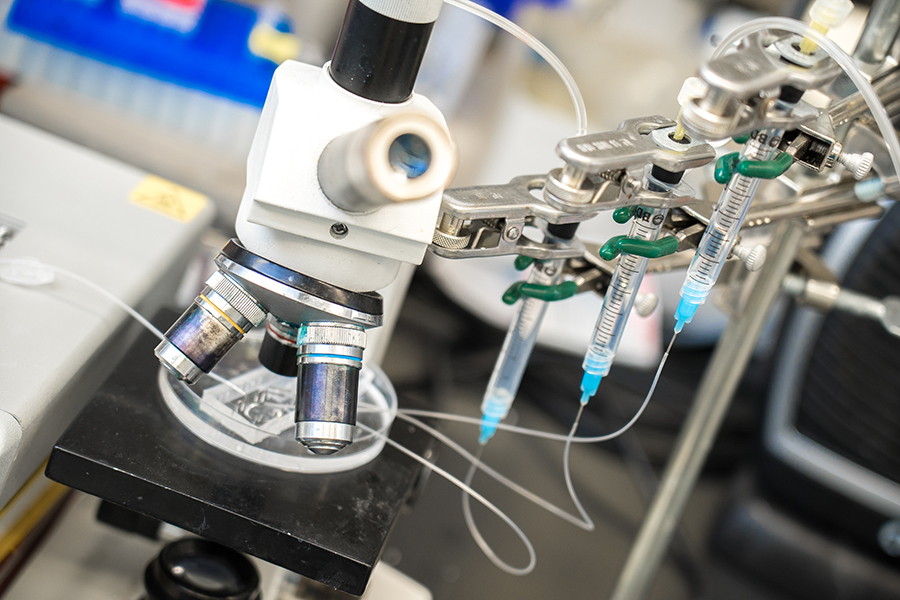
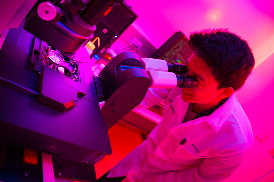
Andrew Richards, a postdoctoral scientist in Zhang's lab, sets up experiments to analyze individual neural cells
One of the projects, awarded $8.7 million over five years from the NIH BRAIN Initiative, is aimed at building a 3D single-cell map for the entire human brain.
“Our ultimate goal is to produce a complete cell atlas of the human brain, including a full catalog of all cell types (a ‘parts list’) and their spatial organization,” Zhang said. “This is a critical step towards understanding the human cognitive machine.”
The new project expands upon Zhang and colleagues’ previous work on mapping diverse populations of neurons in several regions of the brain. It began with a study published in Science that identified 16 neuronal subtypes in six areas of the cerebral cortex. That study was the first large-scale mapping of gene activity in the human brain and provided a basis for understanding the diversity of individual brain cells. Later, armed with a new generation of single-cell sequencing methods, the researchers identified 35 additional subtypes of neural as well as glial cells and discovered which subtypes are most susceptible to common risk factors for different brain diseases.
Moving forward, the researchers will be working on constructing a 3D digital map of all cell types in the entire human brain. They are developing more advanced technology that will enable them to isolate and analyze nuclei of individual human brain cells more efficiently, more precisely and at higher resolution. The researchers will also be developing new computational methods to integrate and analyze these data at much larger scales.
Mapping Respiratory and Urinary Organs
Zhang and colleagues will apply these single-cell sequencing and imaging techniques to build reference maps for organs in the respiratory and urinary systems, including the lungs, kidneys, bladder and ureter. This project was awarded $5.3 million over four years from the NIH’s Human BioMolecular Atlas Program (HuBMAP). It is part of a larger research community effort called the Human Cell Atlas, which aims to “map the adult human body at the level of individual cells.”
The team’s interest in mapping the respiratory and urinary organs stems from their role as so-called “barrier organs”—organs that are responsible for letting in and maintaining the balance of vital substances (oxygen, fluids, electrolytes, etc.) and for eliminating toxic substances from the body.
“Just like what we’re doing with mapping the human brain, we can use this technology to understand what a normal lung, kidney and bladder should look like and how it should function down to the resolution of a single cell. We can then begin to understand the basis of injury and repair of the body’s principal barriers,” said Zhang.
Co-investigators on the BRAIN Initiative project are Jerold Chun, professor of the Degenerative Diseases program and senior vice president of Neuroscience Drug Discovery at Sanford Burnham Prebys Medical Discovery Institute; and Peter Kharchenko, associate professor of biomedical informatics at Harvard Medical School.
Co-investigators on the HuBMAP project are James Hagood, professor of pediatrics at University of North Carolina at Chapel Hill; Sanjay Jain, associate professor of medicine, division of nephrology at Washington University School of Medicine in St. Louis; Peter Kharchenko, associate professor of biomedical informatics at Harvard Medical School; and Xin Sun, professor of pediatrics at the UC San Diego Division of Biological Sciences.
Share This:
You May Also Like


Stay in the Know
Keep up with all the latest from UC San Diego. Subscribe to the newsletter today.